|
|
|
 |
|
|
 |
|
|
|
|
|
|
Living Organisms and the Genetic Code
Asa Packer Elementary
School, Academic Year 2010-2011
Grade 3 |
|
|
|
|
| |
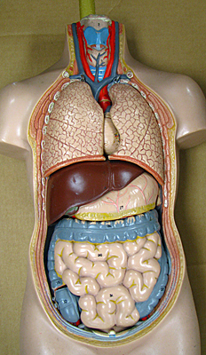
In order to help us with our science unit on Human Anatomy, Dr. DeLeo provided us with a full sized model of the human skeleton. We also got to use a model of the human torso, with organs that came out, and a model of the human heart and brain. We could take the brain apart and put it back together.
We had a lot of fun with the skeleton, called Mr. Bones. He was very nice to us!
|
|
|
|
|
|
|
|
|
|
|
|
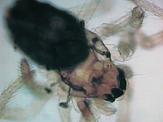
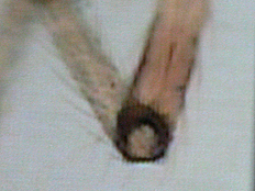
Dr. DeLeo brought a powerful microscope that was connected to a television camera. He placed different things under the microscope and projected the picture on a screen. Dr. DeLeo placed dead bugs - a fly and a spider - under the microscope so they would look real big. It was really scary looking. We are sure glad that real bugs are not that big! Notice the spiky hairs on the photo below on the left. The middle photo shows a spider. The right photo shows a close-up of a joint. We knew it was a joint because we have been studying that in science! |
|
|
|
|
|
|
|
Dr. DeLeo proceeded to place smaller and smaller things under the microscope. |
|
|
|
| |
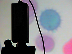
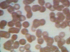
| The photo just to the right shows pollen grains. They are not really that color; they are dyed to make them easier to see. The photo on the far right shows human blood cells. We guessed that one! |
|
|
|
|
|
|
|
| |
|
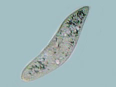
A single cell organism that can be found in pond water is called a paramecium. They are so small that you need a microscope to see them. Dr. DeLeo placed live paramecia under the microscope for us to see. They swim around in a drop of water by moving little hairs along the outside of their bodies. Just think of it - A human being has over 10,000,000,000,000 (ten trillion) cells, but a paramecium has just one!
|
|
|
|
|
|
| |
|
|
| Check out the VIDEOS on the left and right by clicking on the play buttons! The video on the left shows us trying to follow a paramecium that is moving really fast. On the right, we see a paramecium at a higher magnification. |
|
|
|
|
|
|
|
|
|
We learned that each cell can make other cells. A cell does that by dividing into two cells, each the same as the original. That's how we get bigger, and how our body heals itself when we get hurt. Every cell in any plant or animal contains instructions for making that plant or animal. So, every time a cell divides into two cells, the dividing cell has to make two copies of these instructions, one for each of the new cells. The instructions are contained in what are called chromosomes. We call these instructions the "genetic code." |
|
|
|
| |
|
|
|
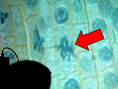
The picture on the left shows the cells in a plant. Plant cells are kind of boxy looking. The circles in the center of the cells are the cell nuclei, and they contain the chromosomes. Some of the cells in the picture are in the process of dividing into two cells. The red arrow points to a cell which is dividing into two cells, and the stringy things are the chromosomes going into both of the new cells! |
|
|
|
| |
|
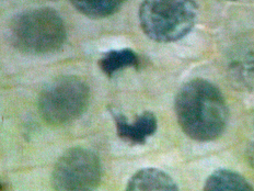
We helped Dr. DeLeo find a really great cell! This one, shown on the right, has just about finished dividing, giving us two cells where before we had only one!
I'll bet Professor Kuchka from Lehigh's Biology Department could even tell us the name of this phase of cell division. Incidentally, Professor Kuchka gave Dr. DeLeo the idea for the next activity! |
|
|
|
|
|
|
|
Dr. DeLeo spoke to us about DNA and the genetic code. The genetic code uses four different chemicals. One ordering makes someone who has blue eyes, another brown eyes. One order makes a human being, another makes an artichoke.
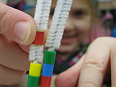
DNA is called a "double helix," a "helix" because it twists like a spiral staircase, and "double" because it is a pair of helixes. The four chemicals have names, but they can be abbreviated by the first letters, A, T, C, and G. The two strands making up the double helix are side-by-side. If there is an "A" on the left, then there will always be a "T" on the right. So, "A" goes with "T" (A-T). Similarly, "C" goes with "G" (C-G). This pairing makes it possible for our cells to divide and make exact duplicates. When a cell is about to divide, the strands come apart, and each one picks up just the right partners to make two new, identical double helixes, one in each of the two cells. |
|
|
|
| |
|
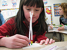
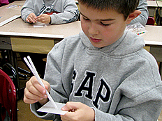
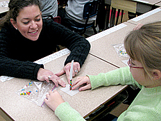
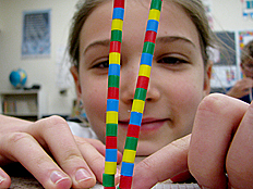
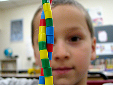
Dr. DeLeo helped us to understand this by giving us beads in fours colors, representing A, T, C, and G. We used a pipe cleaner to put together our make believe DNA. Here are pictures of us putting together our DNA models. We can place any of the four colors in any order along one of the strands. However, where ever there is a blue bead on one side, we must have a yellow bead on the other. And, where ever there is a red bead on one side, we must have a green bead on the other.
|
|
|
|
|
|
|
|
|
|
|
|
|
|
In addition to the DNA models, we each got a cool placemat on human anatomy. We love science! |
|
|
|
|
|
|
|
|
I hope you have enjoyed this web presentation as much as we enjoyed sharing the actual learning experience with your son or daughter. Although we have endeavored to exclude photographs where permission has been denied, it is possible for errors to occur. If you would like us to remove a photograph of your son or daughter for any reason, please send me an e-mail message at lgd0@lehigh.edu or call me at 610-758-3413, and we will remove it promptly. Please note that we will never associate a child's full or last name with a photograph except in circumstances where special permission was explicitly provided. Thank you. Gary DeLeo. |
|
|
|
|
|
|
|
|


Sm.jpg)